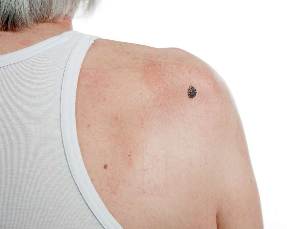
What Does Atopic Dermatitis Rash Look Like? Take A Look
Atopic dermatitis is more than just a common skin condition; it's a chronic affliction impacting millions of lives globally. Characterized by inflamed, itchy skin, this condition goes beyond physical discomfort, affecting emotional well-being and quality of life.

Identifying the Visual Characteristics of Atopic Dermatitis
Atopic dermatitis presents with several distinctive visual features that help differentiate it from other skin conditions. In its early stages, the skin typically appears red and inflamed, with small bumps or papules that may ooze or weep clear fluid when scratched. The affected skin often develops a scaly, dry texture with visible flaking. One of the most distinctive characteristics is the distribution pattern - atopic dermatitis commonly appears in the skin folds, particularly at the elbows, behind the knees, on the neck, and around the eyes and ears. In infants, the rash frequently appears on the cheeks, scalp, and extensor surfaces of the limbs. The appearance can vary significantly between individuals and across different age groups, with some cases presenting as mild, barely noticeable dry patches while others manifest as severe, widespread inflammation.
What Does Atopic Dermatitis Look Like Across Different Skin Tones?
Atopic dermatitis presents differently across various skin tones, which can sometimes lead to delayed diagnosis in darker-skinned individuals. In lighter skin tones, the condition typically appears as red or pink patches with clear boundaries. However, in medium to darker skin tones, atopic dermatitis may present as patches that are darker than the surrounding skin (hyperpigmentation) or sometimes lighter (hypopigmentation). The inflammation can appear purple, gray, or ashen rather than bright red. Additionally, the affected areas may develop a papular presentation (small bumps) more commonly in darker skin. The lichenification—thickening of the skin due to chronic scratching—often appears more prominent in darker skin tones, with more defined skin markings and a leathery texture. Understanding these variations is critical for proper identification regardless of skin color.
Atopic Dermatitis Pictures: Common Presentations and Variations
Visual documentation of atopic dermatitis shows considerable variation in its presentation. In infants, atopic dermatitis often manifests as weepy, crusting patches on the face and scalp. During childhood, the rash typically migrates to the flexural areas, particularly the insides of elbows and knees. Adolescents and adults may develop more prominent lichenification—thickened, leathery skin with accentuated skin lines—due to chronic scratching and inflammation. During flare-ups, the affected skin becomes notably more red, swollen, and may develop small fluid-filled vesicles that can burst and form crusts. In chronic stages, the skin may appear darker or lighter than surrounding areas due to post-inflammatory pigment changes. Some individuals develop a pattern called nummular eczema, characterized by coin-shaped lesions, while others experience more diffuse patches without distinct borders.
Atopic Dermatitis Skin Treatment Options and Effects
Effective treatment of atopic dermatitis can significantly alter the appearance of affected skin. Topical corticosteroids remain a first-line treatment, reducing inflammation and associated redness within days of application. For moderate to severe cases, topical calcineurin inhibitors such as tacrolimus and pimecrolimus work by suppressing specific immune responses without the skin-thinning side effects of long-term steroid use. Moisturizers play a crucial role in treatment regimens by restoring the skin barrier and reducing dryness and flaking. Regular application of thick, fragrance-free emollients helps maintain skin hydration and reduces the scaly appearance characteristic of atopic dermatitis. For severe cases, systemic medications like dupilumab target specific parts of the immune system to reduce inflammation throughout the body. Phototherapy, using controlled exposure to UVB light, can also improve skin appearance by reducing inflammation and itching, though results typically require multiple sessions over several weeks.
Managing Atopic Dermatitis Itching and Its Visual Impact
The intense itching associated with atopic dermatitis significantly affects the visual presentation of the condition through the damage caused by scratching. Breaking the itch-scratch cycle is crucial for improving the skin’s appearance. Cold compresses can provide immediate relief while reducing visible redness and inflammation. Anti-itch topical products containing menthol, pramoxine, or colloidal oatmeal temporarily soothe the sensation while reducing the urge to scratch. Oral antihistamines, particularly sedating ones taken at night, help control nighttime scratching that can otherwise lead to visible excoriations and lichenification. Stress management techniques such as meditation, deep breathing, and gentle exercise can reduce stress-induced flares that worsen both itching and visual symptoms. Wet wrap therapy, involving applying medication and moisturizer followed by wet bandages, not only relieves itching but also improves the appearance of severely affected skin by increasing medication absorption and providing a physical barrier against scratching.
Differentiating Atopic Dermatitis from Similar Skin Conditions
Visual differentiation between atopic dermatitis and other similar-looking skin conditions requires attention to specific characteristics. Contact dermatitis, unlike atopic dermatitis, typically has more defined borders and appears in areas that have direct contact with allergens or irritants. Psoriasis presents with thicker, more silvery scales and well-demarcated plaques, compared to the less distinct boundaries of atopic dermatitis patches. Seborrheic dermatitis primarily affects sebum-rich areas like the scalp, face, and chest with yellowish, greasy scales rather than the dry, flaky appearance of atopic dermatitis. Fungal infections like ringworm typically show a more circular pattern with central clearing and raised borders, unlike the more diffuse pattern of atopic dermatitis. When distinguishing between these conditions proves difficult based on visual inspection alone, dermatologists may perform skin scrapings, patch tests, or biopsies to confirm the diagnosis and guide appropriate treatment.
This article is for informational purposes only and should not be considered medical advice. Please consult a qualified healthcare professional for personalized guidance and treatment.


